Understanding Technique Factors
X- ray technique factors are made up of three variables:
1. Kilovolts (kV) - Controlling the penetration power of the x-ray.
2. Milliamps (mA) - Controlling the volume of x-ray.
3. Time (usually noted in seconds or milliseconds) or pulses (seen in older models) - Controlling the volume of x-ray

Other Items to Consider:
-
Patient Size - a 250 lb adult is almost certain to have denser tissue in the oral-maxillofacial region than that of a 70 lb child.
-
Patient Age - tissue densities will vary between patient ages. Children and elderly patients are more likely to have a lower density than adults.
-
Patient Health - the effects of certain illnesses such as osteoporosis may reduce tissue density.
-
The region within the Oral Cavity - the region around the mandibular anterior teeth has a lower tissue density than around maxillary molars.
The greater the tissue density, the higher the technique factors required to penetrate the tissue and provide satisfactory image quality.
Recognizing Proper Technique Factors
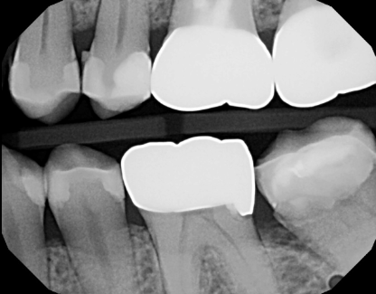
Under Exposed


The above image exhibits slight under-exposure, noticeable through the diminished contrast in the tooth crowns, the gray regions between interproximal spaces and behind molars, and the presence of the positioning tab in the interproximal area. Although under-exposed images can be adjusted for brightness using software, it may not fully restore contrast. It's important to note that this image is also deemed unacceptable due to poor positioning, evident in the contact overlap.
Correct Exposure
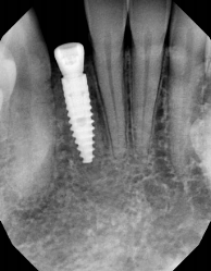
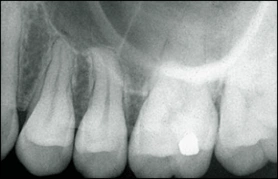
The image above is appropriately exposed, with clear visibility of the apices, DEJ (Dentin-Enamel Junction), and bone details.
Over Exposed

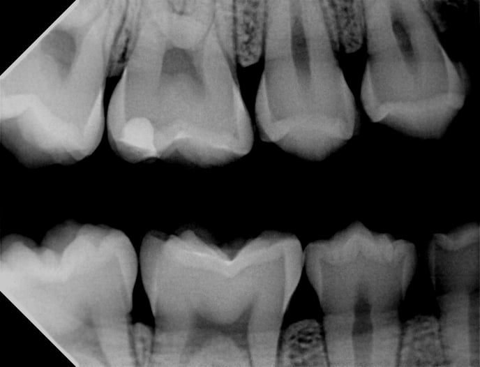
The image displayed above is over-exposed, evident in its overall darkness, particularly noticeable in the cervical burnout on the bicuspids. Adhering to the ALARA (As Low As Reasonably Achievable) principle, it is advisable to reduce the technique factors such as exposure time or pulses to achieve acceptable imaging quality.
Other Considerations
The distance between the X-ray head and the sensor plays a crucial role in image quality. When the X-ray head is farther from the sensor, the amount of radiation reaching the sensor decreases. To ensure consistent imaging, it is recommended to position the cone of the X-ray generator as close to the patient's cheek as possible. Ideally, this involves sliding the ring of the positioning device as close as possible and aligning the cone against the ring.
Overlapping X-rays
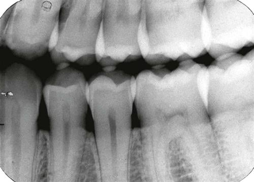
Overlapping X-rays occur when there is unintended superimposition of images on a radiograph. This can lead to difficulties in interpretation and may obscure important details. To avoid overlapping X-rays, it is crucial to ensure proper positioning of the X-ray equipment and the patient. Additionally, employing appropriate techniques, such as using positioning devices and aligning the cone accurately, can help minimize the risk of overlapping and contribute to clear and accurate radiographic images.
Cone Cut X-Rays
-jpg.jpeg?width=240&height=200&name=download%20(1)-jpg.jpeg)
"Cone cut X-rays" refer to radiographs where the X-ray beam does not fully cover the imaging receptor or sensor. This can result in a portion of the image being cut off or incomplete, resembling a "cut" by the X-ray cone.
To prevent cone cuts, it's important to ensure proper alignment and positioning of the X-ray cone with the imaging receptor. This includes verifying that the X-ray cone adequately covers the entire area of interest and that it is centered appropriately. Proper training and adherence to positioning guidelines can help minimize cone cuts and ensure the production of accurate and diagnostically useful X-ray images.
Elongated & Foreshortened X-Rays
Elongated X-Rays:
-
Caused by misalignment or angulation of the X-ray beam, resulting in stretched appearance. Correct by ensuring proper alignment and perpendicular central ray.

Foreshortened X-Rays:
-
Occurs when the X-ray beam is not parallel to the structure, causing a shortened appearance. Prevent by aligning the beam parallel to the structure's long axis and ensuring correct patient and equipment positioning.
|
Principles |
Outcomes |
|---|---|
|
Improves image resolution |
|
Improves image sharpness |
|
Improves image sharpness |
|
Improves anatomic accuracy |
|
Improves anatomic accuracy |
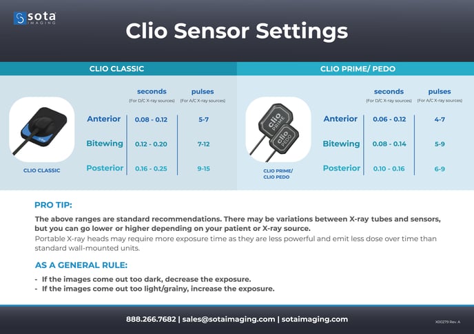
Clio Sensor Settings
-1.png?height=120&name=SotaCloudLogo_LightBG%20(1)-1.png)